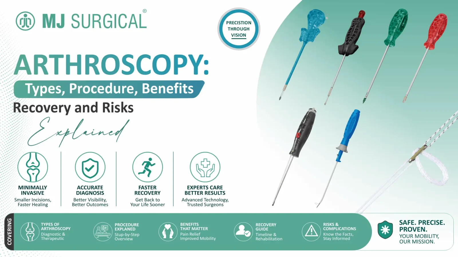
Arthroscopy has quietly become one of the most common ways orthopedic surgeons look inside a joint without opening it up. Instead of a long incision, the surgeon works through a few small cuts, guided by a camera small enough to slip into the joint space itself. What started decades ago as mainly a diagnostic tool has grown into a full surgical technique used to treat torn cartilage, damaged ligaments, and a long list of other joint problems.
If you’ve been told you might need arthroscopy — or you’re a hospital, clinic, or distributor trying to understand the procedure and the instruments behind it — this guide breaks down what arthroscopy actually involves: how it works, where it’s used, what recovery looks like, and what can go wrong. It closes with a look at the instruments that make the procedure possible and where to source them.
What is Arthroscopy?
Arthroscopy is a minimally invasive surgical technique that lets a surgeon examine and treat problems inside a joint through very small incisions. The key instrument is the arthroscope — a narrow tube fitted with a light source and a tiny camera. Once inserted through a small opening (called a portal), it sends a magnified, real-time image of the joint’s interior to a monitor in the operating room.
If the surgeon spots something that needs fixing — a torn meniscus, frayed cartilage, a loose fragment of bone or tissue — additional slim instruments can be passed through nearby portals to treat it during the same procedure. This is what separates arthroscopy from a purely diagnostic scan: in many
cases, the surgeon can look and treat in one sitting, without the larger incision that traditional open surgery requires.
While knee and shoulder arthroscopy are the most familiar examples, the technique is also used on the hip, ankle, elbow, and wrist, with the exact approach shaped by the joint’s anatomy.
Why Do Surgeons Recommend Arthroscopy?
Arthroscopy usually enters the conversation when a joint problem needs a closer look than an X-ray or MRI can provide — or when non-surgical treatment hasn’t resolved persistent pain, swelling, locking, catching, or a feeling of instability in the joint.
Depending on the joint and the underlying issue, arthroscopy may be used to:
- Directly examine structures inside a joint that imaging alone can’t fully assess
- Evaluate and, where appropriate, treat certain sports-related injuries
- Repair or trim damaged meniscus tissue in the knee
- Assess or reconstruct selected ligament injuries
- Address specific cartilage damage
- Remove loose bone or cartilage fragments floating within the joint
- Investigate unexplained joint instability
- Treat certain labral tears in the shoulder or hip
Not every joint problem is a candidate for arthroscopy, though. The decision always comes down to the specific diagnosis, imaging findings, the patient’s overall health, and what the surgeon expects the procedure to actually achieve.
Types of Arthroscopy
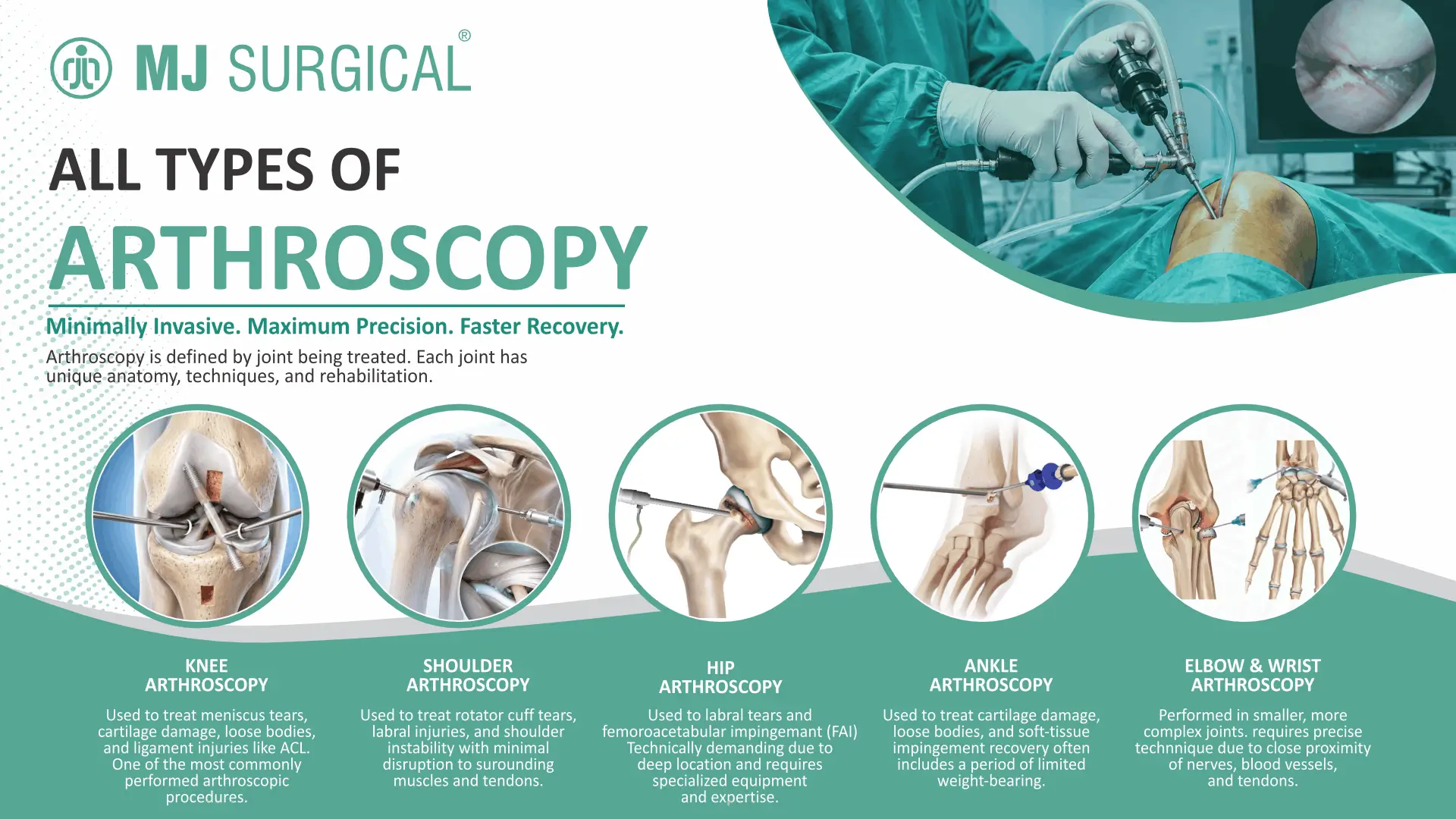
The “type” of arthroscopy is really defined by which joint is being treated, since each joint brings its own anatomy, portal placement, instrumentation, and rehabilitation timeline.
Knee Arthroscopy
Knee arthroscopy is one of the most commonly performed forms of arthroscopic surgery. It gives surgeons a direct view of the meniscus, cartilage surfaces, and ligaments.Depending on what’s found, treatment can range from trimming or repairing torn meniscal tissue to removing loose fragments or reconstructing a torn ligament such as the ACL. Plenty of knee problems still don’t need arthroscopy at all — it comes down to the specific diagnosis.
Shoulder Arthroscopy
Shoulder arthroscopy is commonly used to investigate and treat rotator cuff tears, labral injuries, and issues affecting shoulder stability. Because the shoulder has such a wide range of motion and a comparatively shallow joint socket, arthroscopic techniques are especially useful for repairing soft-tissue damage without disturbing the surrounding muscle and tendon structures more than necessary.
Hip Arthroscopy
The hip sits deep beneath a thick layer of muscle, which makes hip arthroscopy technically more demanding than knee or shoulder procedures. It’s typically considered for conditions like labral tears or femoroacetabular impingement (FAI), and it calls for specialized positioning equipment, longer instruments, and surgeons experienced specifically in hip arthroscopic technique.
Ankle Arthroscopy
Ankle arthroscopy addresses issues such as cartilage damage, loose bodies, and soft-tissue impingement within the joint. Because the ankle is a relatively tight, weight-bearing joint, recovery often includes a period of limited weight-bearing, with the exact restrictions depending on what was treated.
Elbow and Wrist Arthroscopy
These are smaller, more anatomically crowded joints, with major nerves, blood vessels, and tendons running close to the surgical field. Elbow and wrist arthroscopy demand precise portal placement and instrumentation suited to that tight anatomy, which is why they’re generally performed by surgeons with specific experience in these joints.
How is Arthroscopy Performed?
The exact steps differ depending on the joint and diagnosis, but most arthroscopic procedures follow a similar general sequence.
1. Preoperative Assessment
Before the day of surgery, the surgical team reviews the patient’s symptoms, medical history, physical exam findings, and imaging. The surgeon walks the patient through what the procedure involves, what recovery typically looks like, possible complications, and any alternatives worth considering.
2. Anesthesia
The anesthesia approach — general, regional, or local — is chosen based on the joint involved, the complexity of the planned procedure, and the patient’s overall health. This decision is made jointly by the surgical and anesthesia teams ahead of the operation.
3. Creating the Portals
The surgeon makes one or more small incisions, known as portals, around the joint. The arthroscope goes in through one; additional portals allow other instruments to be introduced as needed.
4. Examining the Joint
With the arthroscope in place, the surgeon gets a magnified, illuminated view of the joint’s interior on a monitor, allowing a close inspection of the structures involved before deciding on next steps.
5. Performing the Treatment
If a treatable problem is confirmed, the surgeon introduces additional slim instruments through the other portals — to trim damaged tissue, remove loose fragments, repair a structure, or complete a reconstruction, depending on what the diagnosis calls for.
6. Closing Up
Once treatment is complete, the arthroscope and instruments are withdrawn, and the small portal incisions are closed with sutures, adhesive strips, or similar methods. Many arthroscopy procedures are done as same-day surgery, though some cases call for an overnight stay.
Benefits of Arthroscopy
Compared with traditional open surgery, arthroscopy offers several potential advantages when it’s the right fit for the condition being treated:
- Smaller incisions than most open surgical approaches
- Direct, magnified visualization of structures inside the joint
- The ability to diagnose and treat certain conditions in the same operation
- Generally less disruption to surrounding muscle and soft tissue
- Often a quicker recovery than a comparable open procedure
- A technique applicable across a wide range of orthopedic and sports injuries
That said, minimally invasive doesn’t automatically mean better in every case. Some injuries genuinely need an open surgical approach, and the right call always depends on the diagnosis, the joint involved, and the surgeon’s clinical judgment.
Arthroscopy Recovery: What to Expect
Recovery timelines vary a lot from one patient to the next, largely because “arthroscopy” covers everything from a quick diagnostic look to a full ligament reconstruction. A simple procedure might mean a return to light activity within days; a repair or reconstruction can mean weeks or months of structured rehabilitation.
During recovery, patients are typically advised to:
- Protect the treated joint as instructed by the surgical team
- Manage pain and swelling according to the post-op plan
- Take medications exactly as prescribed
- Follow any weight-bearing or movement restrictions
- Complete recommended exercises or physiotherapy sessions
- Increase activity gradually rather than rushing back
- Attend all scheduled follow-up appointments
It’s worth resisting the urge to compare your recovery timeline to someone else’s — the joint, the procedure, and each person’s healing all play a role. The rehabilitation plan from your own surgical team is the one that matters.
Possible Risks and Complications of Arthroscopy
Small incisions don’t mean zero risk — arthroscopy is still surgery. Potential complications include:
- Infection
- Bleeding or blood collection within the joint
- Blood clots
- Injury to nearby nerves or blood vessels
- Damage to structures within the joint
- Joint stiffness or persistent pain
- Anesthesia-related complications
- The possibility of needing further treatment or surgery
Most patients get through arthroscopy without serious complications, but the level of risk depends on the joint, the specific procedure, and the patient’s individual health background. Anyone who develops severe or worsening pain, significant swelling or redness, fever, unusual discharge from the incision, or numbness and tingling after surgery should contact their surgical team promptly.
Arthroscopy Instruments and Equipment
Behind every arthroscopy procedure is a set of purpose-built instruments designed to visualize, grasp, cut, repair, and access tissue through portals just a few millimeters wide.
Arthroscopes
The arthroscope is the core visualization tool — a rigid optical instrument connected to a camera and light source that projects the inside of the joint onto a monitor in real time.
Graspers and Probes
Graspers hold and retrieve tissue or loose fragments during the procedure, while probes let the surgeon physically assess the condition of cartilage, ligaments, and other structures inside the joint.
Cutting and Shaving Instruments
Specialized punches, shavers, and cutting instruments are used when damaged tissue needs to be trimmed, removed, or reshaped.
Tissue Repair and Fixation Instruments
Certain arthroscopic procedures call for dedicated repair instruments — suture passers, anchors, and related devices — chosen according to the specific structure being repaired.
Cannulas and Accessories
Cannulas keep portal access controlled and stable, making it easier to pass instruments in and out of the joint throughout the procedure.
Choosing the Right Arthroscopy Instruments
For hospitals, surgeons, and distributors building or updating an arthroscopy set, the selection process usually comes down to a handful of practical factors:
- Intended surgical application and joint
- Instrument design, ergonomics, and precision
- Material quality and durability
- Compatibility with the existing arthroscopy system
- Cleaning and sterilization requirements
- Manufacturing quality and regulatory compliance
- Supplier reliability and product availability
Every facility should weigh these against its own clinical caseload, sterilization workflow, and procurement requirements before finalizing a purchase.
Arthroscopy vs. Open Surgery
The core difference comes down to access. Arthroscopy works through a handful of small portals; open surgery uses a larger incision to expose the surgical site directly. Smaller incisions generally mean less disruption to surrounding tissue for procedures where arthroscopy is appropriate — but it isn’t a universal upgrade. Certain complex injuries genuinely need the direct access that open surgery provides. The right approach depends on the diagnosis, anatomy, and the outcome the surgeon is aiming for.
Arthroscopy vs. Arthroplasty
These two terms get mixed up often, but they describe very different procedures. Arthroscopy uses a camera and small instruments to examine or treat structures inside an existing joint. Arthroplasty, on the other hand, involves reconstructing or replacing a damaged joint or joint surface altogether — total knee and hip replacements being the most familiar examples. Put simply: arthroscopy tends to preserve and repair the joint you have, while arthroplasty rebuilds it. Which one applies depends on how much joint damage is present and what the patient needs functionally.
Why Choose MJ Surgical for Arthroscopy Instruments
MJ Surgical manufactures and supplies orthopedic surgical instruments and implants, including products developed for arthroscopy applications, from our facility in Ahmedabad, India. Our arthroscopy-related products are designed with attention to precision, ergonomics, and material quality, and are manufactured under quality-controlled processes intended to meet the practical demands of orthopedic surgical teams.
Whether you’re a hospital building out a minimally invasive surgery program, a distributor sourcing for multiple facilities, or a surgeon evaluating new instrumentation, our team can walk you through product specifications, compatibility, and availability.
Explore MJ Surgical’s Arthroscopy Product Range →
Frequently Asked Questions About Arthroscopy
What is arthroscopy used for?
Arthroscopy is used to examine and, where appropriate, treat problems inside a joint — including certain meniscus, cartilage, ligament, and labral conditions.
Is arthroscopy a major surgery?
It’s classified as minimally invasive because it uses small portals rather than a large incision, but it’s still surgery. Complexity ranges from straightforward diagnostic procedures to more involved repairs and reconstructions.
How long does an arthroscopy procedure take?
It depends on the joint and the treatment involved — some procedures are relatively quick, while more complex repairs can take considerably longer.
How long is recovery after arthroscopy?
Recovery ranges from a few days for minor procedures to several weeks or months for repairs and reconstructions, depending on the joint and the extent of treatment.
Is arthroscopy painful?
Some pain, swelling, and stiffness after surgery is normal and varies by procedure and individual. The surgical team provides a specific plan for pain management and wound care.
Is arthroscopy better than open surgery?
Not automatically. It can be advantageous for certain conditions, but some injuries are better suited to an open surgical approach. The decision rests on the diagnosis and the surgeon’s assessment.
Which joints can be treated with arthroscopy?
Most commonly the knee and shoulder, though hip, ankle, elbow, and wrist arthroscopy are also performed for select conditions.
Conclusion
Arthroscopy has changed how orthopedic surgeons diagnose and treat joint problems — offering a way to look inside a joint and, often, fix what’s wrong, all through a few small incisions. Knee and shoulder arthroscopy remain the most common applications, but the technique extends to the hip, ankle, elbow, and wrist when the situation calls for it.
For hospitals, surgeons, and distributors, having the right arthroscopy instruments on hand is just as important as the surgical technique itself. MJ Surgical supplies orthopedic instruments and implants built for the demands of modern minimally invasive surgery.
About the Author
MJ Surgical Clinical & Product Team.
This article was prepared by the MJ Surgical Clinical & Product Team, drawing on established medical and orthopedic education resources. The content is intended for educational and informational purposes.
References
This article draws on publicly available patient-education resources published by recognized medical authorities:
1. American Academy of Orthopaedic Surgeons (AAOS) — OrthoInfo: Arthroscopy — the peer-reviewed patient information portal of the leading U.S. professional association for orthopaedic surgeons.
2. National Health Service (NHS, UK) — Arthroscopy — the United Kingdom’s public national healthcare service.
Disclaimer
This information is for educational purposes only and not a substitute for professional medical advice. Always consult your doctor for medical concerns.